- Home
- Knee Joint Anatomy
- Knee Bones
Knee Bones
Written By: Chloe Wilson, BSc(Hons) Physiotherapy
Reviewed by: KPE Medical Review Board
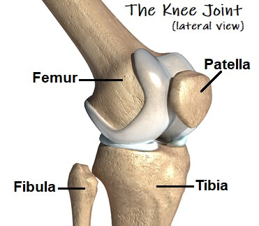
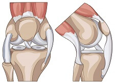
There are three knee bones that make up the knee joint:
- Femur: aka thigh bone runs from the hip to the knee
- Tibia: aka shin bone runs from the knee to the ankle, and
- Patella: aka kneecap is the small bone at the front of the knee.
The two largest knee bones, the femur and the tibia, join together to form what is known as the tibiofemoral joint, and at the front of the knee the kneecap rests in a groove on the front of the femur, known as the patellofemoral joint.
Another bone, the fibula, is found on the outside of the leg. It is not directly part of the knee joint, but many of the important structures of the knee attach to the fibula.
The bones of the knee are all lined with cartilage and whole joint is surrounded by a joint capsule.
The Knee Bones
Here we will look at each the different knee bones and their associated structures and how they work. We will also go on to look at what can go wrong with them and how to treat any problems that may arise.
1. Femur (Thigh Bone)
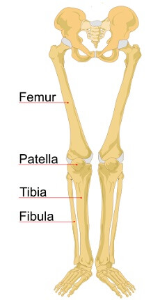
The femur is the longest bone in the body found at the top of the leg, sitting between the pelvis/hip and the knee. It measures approximately one quarter of your height!
At the hip, the femur has a round shaped head which articulates with the part of the pelvis known as the acetabulum - a deep socket that provides lots of stability.
At the knee, the femur articulates with the shin bone (tibia) and the knee cap (patella).
On the bottom end of the femur are two round knobs known as the femoral condyles which rest on top of the shin bone. Between these knobs is a shallow groove called the patella groove. The knee cap rests in this and glides freely up and down the groove as you bend and straighten your leg.
2. Tibia (Shin Bone)
The tibia is the second longest bone in the human body that is found between the knee and the foot. It has a long, triangular shape and bears a majority of the body's weight when you are standing.
The top surface of the tibia is essentially flat and is known as the tibial plateau. The femur sits on this flat area.
The top of the tibia is lined with an extra layer of cartilage, known as the meniscus. Damage to this cartilage is a common cause of knee pain and may be due to a cartilage tear or arthritis.
About 2 cm below the joint line on the front of the tibia is a bony prominence known as the tibial tuberosity. This is where the patellar tendon attaches to join the quads muscles to the tibia. Tightness in the quads muscles can pull on the tibial tuberosity causing it to lay down extra bone as a protective measure which leads to the formation of a hard lump on the front of the shin bone. This is known as Osgood Schlatters Disease and is common in adolecents, particularly following a growth spurt.
The bottom end of the tibia widens into a bony prominence called the medial malleolus, forming the inner side of the ankle joint.
3. Patella (Kneecap)
The patella is a small upside down triangle-shaped bone found at the front of the knee. The patella bone sits inside the tendon of the thigh muscles, the quadriceps, at the front of the knee resting in the patellar groove of the femur.
Not only does the patella protect the other knee bones, it also plays an important role in knee stability and increases the leverage and mechanical function of the quadriceps muscles, so the knee can function efficiently.
A great deal of pressure goes through the kneecap and it is therefore lined with the thickest layer of cartilage in the body.
Common problems around the patella include:
- Chondromalacia Patella: damage to the cartilage
- Housemaids Knee aka prepatellar bursitis
- Patella Alta aka high riding patella
- Patellofemoral Pain Syndrome aka Runners Knee
The most common injuries of the kneecap bone are dislocations or fractures - you can find out more in the kneecap injuries section.
4. Fibula
The fibula is often mistaken as being a knee bone. It is the long, thin bone that runs down the outer side of the lower leg between the knee and foot, and is thinner and smaller than the tibia.
The fibula is not technically part of the knee joint but it is joined to the tibia by muscles so can be involved in various problems in the knee. No weight actually goes through the fibula when you are standing, but it does help to enhance the strength and stability of the lower leg.
Structures Around the Knee Bones
Located around the knee bones are a number of different structures that stabilise and control the joint and allow it to move.
1. Articular Cartilage
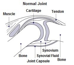
Each of the knee bones is covered in a thin layer of articular cartilage.
This cartilage lines the bones providing some cushioning and allows them to slide smoothly on each other without friction as the leg bends and straightens.
The meniscus is a much thicker specialist layer of cartilage unique to the knee joint. It supplements the articular cartilage to provide extra cushioning to help protect the knee bones from the large forces that go through the joint.
Damage to the meniscus is a frequent cause of knee pain and can be notoriously slow to heal. You can find out all about the causes, symptoms, diagnosis and treatment options in the meniscus tear section.
2. Joint Capsule
The knee joint capsule is like a bag that surrounds the entire joint and attaches to the knee bones.
The joint capsule contains liquid called synovial fluid which lubricates and nourishes the knee joint, like oil in your car engine. This fluid is constantly replenishing itself, especially during movement of the knee.
If you have been sitting or sleeping for long periods, the knee joint capsule can dry out making movements painful initially. As you move the knee, it starts to pump synovial fluid into the joint capsule and the stiffness and pain settle down.
Morning stiffness in the knee is a typical symptom of arthritis, due to the drying up of the synovial fluid. Once you start moving around, the knee starts producing more fluid and the stiffness and pain settles.
Some people have folds in the synovial membrane, known as plica, which can get inflammed, known as plica syndrome, causing a dully achy pain in the knee which gets worse with activity and clicking noises as you bend and straighten the knee.
3. Knee Ligaments
Ligaments are thick, fibrous bands that connect bones. There are two sets of ligaments that attach to the knee bones:
- Cruciate Ligaments: ACL & PCL which sit deep inside the knee joint
- Collateral Ligaments: MCL and LCL which attach to the sides of the knee bones
The knee ligaments play a vital role in providing stability to the knee bones and joints. The knee ligaments are frequently injured in twisting injuries. Overstretching of the ligaments leads to tearing of some of fibres known as a sprain or rupture.
MCL tears are the most common ligament injury, followed by ACL tears. PCL tears and LCL tears are less common. Ligament tears tend to affect knee stability and which can damage the knee bones.
4. Knee Muscles
There are two main sets of muscles that attach to the knee bones and allow the joint to move:
- Quadriceps: Attach at the front of the knee and control knee extension
- Hamstrings: Attach at the back of the knee and control knee flexion
The quadriceps and hamstrings work together to contract and relax to allow smooth, controlled movement at the knee by pulling on the knee bones.
Common Knee Bone Injuries
The knee bones are very strong but are susceptible to damage. The two most common things that can happen are:
- Fractures: where one or more of the knee bones is broken, usually due to a high velocity force through the bone e.g. RTA or fall. Find out more about knee fractures
- Knee Bone Spurs: wear and tear or excessive pressure/friction leads to excess bone production in the knee which results in hard lumps of excess bone forming on parts of the knee. You can find out all about the causes, symptoms and treatment options in the bone spurs in knee section.
What Next?
If you have enjoyed learning about the knee bones and would like to know more about the other structures that make up the knee, visit the knee anatomy guide or choose from the following:
If you are suffering with knee pain and want some help working out what is causing your problem, visit the knee pain diagnosis section.
You may also be interested in the following articles:
Page Last Updated: 18/06/23
Next Review Due: 18/06/25
