- Home
- Knee Joint Anatomy
- Ligaments
Knee Ligaments
Written By: Chloe Wilson, BSc(Hons) Physiotherapy
Reviewed by: KPE Medical Review Board
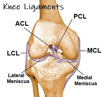
The knee ligaments are one of the most vital components of knee stability and control.
Ligaments are strong, thick fibrous bands, like ropes, that connect bone to bone, provide stability, control movement and prevent injury.
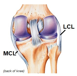
There are two main pairs of ligaments in the knee, the cruciate ligaments in the center of the knee (ACL & PCL) and the collateral ligaments on the sides of the knee (MCL & LCL).
The ligaments of the knee are frequently damaged by sudden twisting movements e.g. changing direction quickly when running, or a force through the knee e.g. a fall or tackle.
After a
ligament injury,
the knee can feel painful, weak and unstable. This may only last a few days, but if left untreated, can persist for many months, so early treatment is key.
Here we will look at the different knee ligaments, where they are located, how they work and how they get injured.
Ligaments Of The Knee
The four main ligaments in the knee are:
- Medial Collateral Ligament: found on the inner side of the knee
- Lateral Collateral Ligament: found on the outer side of the knee
- Anterior Cruciate Ligament: found in the middle of the knee
- Posterior Cruciate Ligament: found in the middle of the knee
The collateral and cruciate ligaments are generally considered to be the main ligaments of the knee but there are a few others that work alongside them, although not everyone has all of them!
Collateral Knee Ligaments
The collateral ligaments are responsible for the sideways stability of the knee and the cruciate knee ligaments are responsible for the forwards and backwards stability of the knee. Both provide stability with twisting movements as well.
1. Medial Collateral Ligament (MCL)
The medial collateral ligament is found on the medial (inner) side of the knee.
It is a broad flat ligament of the knee, approximately 10 cm long, that attaches to the femur and the tibia.
The MCL resists forces through the outer side of the leg that push the knee inwards, known as valgus forces.
The medial collateral ligament gets damaged when the ligament is overstretched and tears, typically from:
- Contact Injury: When there is a force through the outer side of the knee e.g. sporting tackle
- Twisting Injury: Twisting awkwardly, particularly when the foot is fixed e.g. quickly changing direction when running.
MCL tears result in pain and swelling on the inner side of the knee joint and the knee may feel unstable depending on the severity of the injury.
Visit the MCL injuries to find out loads more including treatment and prevention strategies.
2. Lateral Collateral Ligament (LCL)
The lateral collateral ligament, also known as the fibular collateral ligament, is found on the outer side of the knee, connecting the femur to the fibula. It resists forces from the inner side of the knee (known as varus forces) and is the primary restraint against forces that push the knee outwards.
The lateral collateral ligament is much shorter than the medial collateral ligament making it much less common to injure the LCL than the MCL.
Cruciate Knee Ligaments
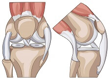
The cruciates are the most important knee ligaments in providing stability of the knee. There are two cruciate ligaments, anterior (ACL) and posterior (PCL).
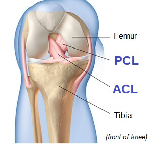
They sit deep inside the middle of the joint attaching to the tibia and femur. They cross over each other, hence their name, and resemble the St Andrews Cross (X).
The cruciate knee ligaments are each about as thick as a pencil and are extremely strong, with a breaking strain of about 60kg.
They get their name from where they attach to the tibia, i.e. the ACL attaches to the anterior (front) surface of the tibia and the PCL to the posterior (back) surface.
The cruciate ligaments job is to control the forwards and backwards movement of the knee joint. They are also important in providing proprioception – the body's ability to know where it is and to make subtle adjustments to maintain balance.
Each knee cruciate ligament is about 2cm long and any force which
stretches it an additional 1.7mm (8% total length) will result in
complete ligament tear.
1. Anterior Cruciate Ligament (ACL)
The anterior cruciate ligament sits deep in the middle of the knee joint. It attaches to the front of the tibia and the back of the femur.
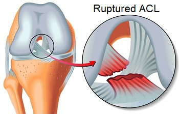
The ACL stops the tibia sliding too far forward in relation to the femur and is the primary structure for proprioception.
The anterior cruciate ligament is commonly injured in sporting activities such as football and skiing, usually from awkward twisting movements, sudden stopping or landing awkwardly.
It can take up to a year to recover from an ACL injury so prevention has become an important factor in sports training.
To find out more visit the ACL injuries section for in-depth information on injuries, surgery and rehab.
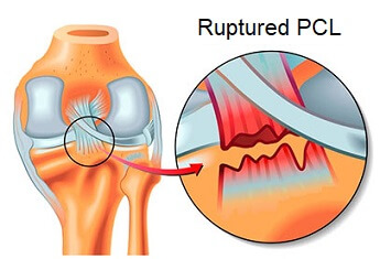
2. Posterior Cruciate Ligament (PCL)
The posterior cruciate ligament also sits deep in the knee joint and it attaches to the back of the tibia and the front of the femur.
The PCL is shorter than the ACL, at 3/5 of the length, but is twice as strong. As a result it is much harder to injure to PCL.
The PCL stops the tibia moving too far back in relation to the femur.
The posterior cruciate ligament is typically damaged by a sudden force through the top of the shin, from a car accident or fall, or by hyperextending the knee.
You can find out loads more about the causes, symptoms, treatment and prevention strategies in the PCL injury section.
Other Knee Ligaments
There are other less well known knee ligaments are:
- Transverse Ligament: joins the medial and lateral meniscus
- Anterior Meniscofemoral Ligament: joins the lateral meniscus to the medial femoral condyle
- Posterior Meniscofemoral Ligament: joins the lateral meniscus to the medial femoral condyle
- Coronary Ligaments: join both menisci to the tibia
- Oblique Popliteal Ligament: attaches to one of the hamstring tendons
- Arcuate Popliteal Ligament: joins the fibula to the tibia and femur
Damage to any of these ligaments can cause pain and inflammation but they are unlikely to result in knee instability unless they are injured at the same time as the collateral or cruciate knee ligaments.
Common Knee Ligament Injuries
Anything that stretches the knee ligaments beyond their elastic limit will cause the ligament to tear. Ligament tears vary from a minor sprain where only a few fibers are torn to major tears where the ligament ruptures completely.
When one or more of the knee ligaments are torn, it can cause real ongoing problems with stability of the knee, so prompt rehab is vital.
There are three different grades of knee ligament injuries depending on the extend of the damage
- Grade 1 Ligament Tear: mild stretching or microscopic tearing of one or more of the knee ligaments, causing minimal instability
- Grade 2 Ligament Tear: partial tears of the ligament leading to increased joint laxity and moderate instability
- Grade 3 Ligament Tear: complete rupture of one of the knee ligaments resulting in significant joint instability
Prompt treatment is vital to reduce pain and inflammation and ongoing rehab helps to reduce the instability at the knee and prevent further injuries.
You can find out more about knee ligament injuries, including the causes, symptoms, treatment and injury prevention in the following articles:
Quiz Time: What is the difference between a ligament and a tendon?
A ligament joins bone to bone whereas a tendon joins bone to muscle.
You may also be interested in the following articles:
- Front Knee Pain
- Pain Behind The Knee
- Inner Knee Pain
- Outer Knee Pain
- Burning Knee Pain
- How To Do Stairs With Knee Pain
- Knee Strengthening Exercises
Page Last Updated: 17/05/23
Next Review Due: 17/05/25
